Hundred genes and many genomic regions have been identified as potentially causative for human neurodevelopmental disorders such as autism, intellectual disability/developmental delay, epilepsy, and schizophrenia (Cooper et al. 2011; Girirajan et al. 2011; O'roak et al. 2012; Fromer et al. 2014; Gilissen et al. 2014; Iossifov et al. 2014). While these studies are invaluable in providing a list of candidate genes, the molecular genetic basis of how disruption of these genes lead to disease is not completely understood (Chakravarti et al. 2013). Moreover, current strategies for functional evaluation of the discovered genes in animal models are limited due to a lack of highly sensitive and quantitative assays. Mouse models have been an impeccable resource for researchers, but functional genetics using mice are expensive, time consuming, and laborious. For decades, Drosophila melanogaster has proven to be a powerful model for genetic studies with about 75% of human disease genes having orthologs in flies (Reiter et al. 2001; Inlow and Restifo 2004). The genetic system and tools developed using Drosophila have provided us with a deeper understanding of cellular and molecular basis of several basic biological processes (St Johnston 2002). With the availability of such tools and high conservation of human disease-associated genes, the past decade has also seen the growth of Drosophila models to study human diseases (Wangler et al. 2015).
The Drosophila compound eye is a simple nervous system consisting of a symmetrical organization of approximately 750 ommatidia (Ready et al. 1976). Two-thirds of the vital genes in the Drosophila genome have been estimated to be required for eye development (Thaker and Kankel 1992). Although some genes are likely to be specific for eye development, other vital genes expressed in the eye are probably required for general cellular processes as well (Thomas and Wassarman 1999). Hence, phenotypic assessment of the eye can be extended to gene functions in other tissues. Since it is a dispensable organ for survival, studies using the fly eye have been used for understanding basic biological processes including cell proliferation and differentiation, neuronal connectivity, apoptosis, and tissue patterning (Karim et al. 1996). Classical studies on Drosophila have established a line of research using the fly eye as an experimental system for studying genetic effects (Ready et al. 1976; Meyerowitz and Kankel 1978; Moses and Rubin 1991; Thaker and Kankel 1992; Kumar 2012). For decades, the fly eye has been used as a system for functional screening of genes and genetic interactions involved in basic cellular processes, neurodevelopment and degeneration, and common complex diseases such as cancer and diabetes (Thomas and Wassarman 1999). We present a computational method for high throughput assessment of eye morphology in fruit flies. Flynotyper provides an accurate and automated method for eye phenotyping. Our algorithm is implemented as a software package called Flynotyper available for free download.
Eye imaging details for phenotyping using Flynotyper:
Eye imaging using bright field microscope: For light microscope imaging of adult eyes, we recommend immobilizing flies by freezing at -80°C and then mounting on blu-tack (Bostik Inc, Wauwatosa, WI). Please note that small errors in ommatidial detection can drastically change the interpretation of phenotypes, and Flynotyper will be more accurate when the fly eye is mounted in the right orientation. Mounting the samples at an angle can lead to incorrect phenotypic scores. We have used Olympus BX53 compound microscope with a LMPlanFL N 10X 0.25 NA air objective (Olympus, Tokyo, Japan), at 0.5X magnification and a z-step size of 12.1μm for imaging. We captured the images using CellSens Dimesion software (Olympus Optical) and then the image slices were stacked using Zerene Stacker (Zerene Systems, USA). Note that a higher quality contrast image would work best for ommatidial detection and obtaining phenotypic scores.
Eye imaging using Scanning Electron Microscope (SEM): Standard protocols for obtaining SEM images from flies can be followed. Flynotyper works best if the eyes are mounted in the right orientation. Manually cropping the region of interest in an SEM image gives a more accurate phenotypic assessment.
References
- Chakravarti, A., A. G. Clark and V. K. Mootha, 2013 Distilling pathophysiology from complex disease genetics. Cell 155: 21-26.
- Cooper, G. M., B. P. Coe, S. Girirajan, J. A. Rosenfeld, T. H. Vu et al., 2011 A copy number variation morbidity map of developmental delay. Nat. Genet. 43: 838-846.
- Fromer, M., A. J. Pocklington, D. H. Kavanagh, H. J. Williams, S. Dwyer et al., 2014 De novo mutations in schizophrenia implicate synaptic networks. Nature 506: 179-184.
- Gilissen, C., J. Y. Hehir-Kwa, D. T. Thung, M. van de Vorst, B. W. van Bon et al., 2014 Genome sequencing identifies major causes of severe intellectual disability. Nature 511: 344-347.
- Girirajan, S., C. D. Campbell and E. E. Eichler, 2011 Human copy number variation and complex genetic disease. Annu. Rev. Genet. 45: 203-226.
- Inlow, J. K., and L. L. Restifo, 2004 Molecular and comparative genetics of mental retardation. Genetics 166: 835-881.
- Iossifov, I., B. J. O'Roak, S. J. Sanders, M. Ronemus, N. Krumm et al., 2014 The contribution of de novo coding mutations to autism spectrum disorder. Nature 515: 216-221.
- Karim, F. D., H. C. Chang, M. Therrien, D. A. Wassarman, T. Laverty et al., 1996 A screen for genes that function downstream of Ras1 during Drosophila eye development. Genetics 143: 315-329.
- Kumar, J. P., 2012 Building an ommatidium one cell at a time. Dev. Dyn. 241: 136-149.
- Meyerowitz, E. M., and D. R. Kankel, 1978 A genetic analysis of visual system development in Drosophilia melanogaster. Dev. Biol. 62: 112-142.
- Moses, K., and G. M. Rubin, 1991 Glass encodes a site-specific DNA-binding protein that is regulated in response to positional signals in the developing Drosophila eye. Genes Dev. 5: 583-593.
- O'Roak, B. J., L. Vives, S. Girirajan, E. Karakoc, N. Krumm et al., 2012 Sporadic autism exomes reveal a highly interconnected protein network of de novo mutations. Nature 485: 246-250.
- Ready, D. F., T. E. Hanson and S. Benzer, 1976 Development of the Drosophila retina, a neurocrystalline lattice. Dev. Biol. 53: 217-240.
- Reiter, L. T., L. Potocki, S. Chien, M. Gribskov and E. Bier, 2001 A systematic analysis of human disease- associated gene sequences in Drosophila melanogaster. Genome Res. 11: 1114-1125.
- St Johnston, D., 2002 The art and design of genetic screens: Drosophila melanogaster. Nat. Rev. Genet. 3: 176- 188.
- Thaker, H. M., and D. R. Kankel, 1992 Mosaic analysis gives an estimate of the extent of genomic involvement in the development of the visual system in Drosophila melanogaster. Genetics 131: 883-894.
- Thomas, B. J., and D. A. Wassarman, 1999 A fly's eye view of biology. Trends Genet. 15: 184-190.
- Wangler, M. F., S. Yamamoto and H. J. Bellen, 2015 Fruit Flies in Biomedical Research. Genetics.
System Requirements
Flynotyper requires the OpenCV4 library for image processing and the wxWidgets 3.2 libraries to run the desktop application. To install these, your system will also need GCC 11.4.0 or higher, GTK 3.X, and CMake 2.6 or higher. Please install OpenCV4 and its dependencies by following the instructions in the "Installation" section.
Installation
Install OpenCV4
Make sure you have GCC and CMake installed on your system. To ensure you download the latest version of OpenCV, use the -b flag in the git command:
$ cd ~<my_working_directory> $git clone -b 4.x https://github.com/Itseez/opencv.git
Build OpenCV4
$ cd ~<my_working_directory> $ mkdir build
$ cd build
$ cmake -D CMAKE_BUILD_TYPE=RELEASE -D CMAKE_INSTALL_PREFIX=/usr/local ..
$ make
$ sudo make install
Install wxWidgets
You can install the binaries for wxWidgets here
Build wxWidgets
For more information on how to build wxWidgets, go to this tutorial created by the developers of wxWidgets.
$ cd wxWidgets-3.2.x
$ mkdir gtk-build
$ cd gtk-build
$ ../configure
$ make -j3
$ sudo make install
$ sudo ldconfig
Install Flynotyper 2.0
Linux
Download the source code from our GitHub page.
$ cd flynotyper-desktop-application
$ make
This will create an executable labeled "desktop_app"
Mac
Download the source code listed above. Then, run the following:
$ export DYLD_LIBRARY_PATH=/usr/local/lib:$DYLD_LIBRARY_PATH
From there, run the same commands as you would on Linux:
$ cd flynotyper-desktop-application
$ make
This will create an executable labeled "desktop_app"
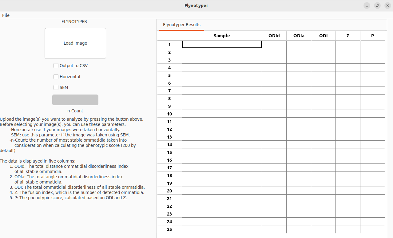
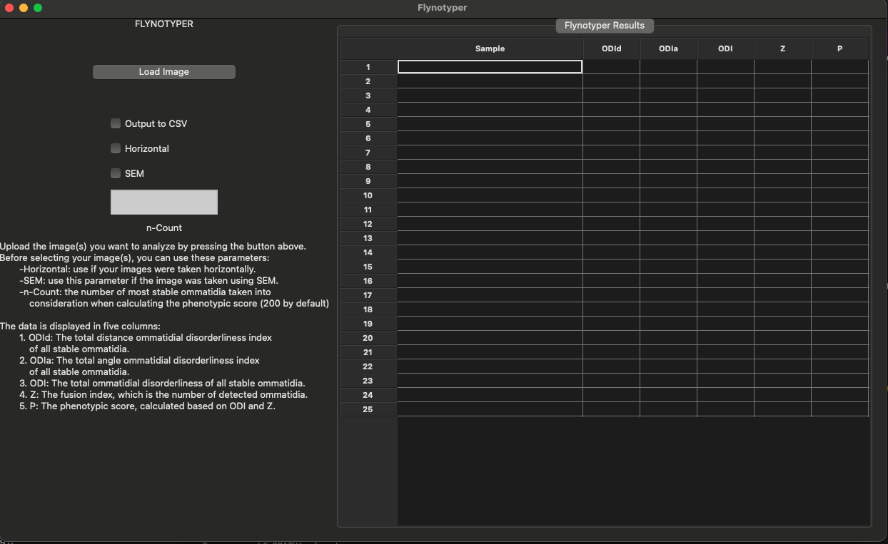
Usage
To open the application, either click on the executable or run it in the command line.
Flags
- Output to CSV: if you want to output your data into a .csv file, use this flag.
- SEM: use this flag if the image was taken using SEM.
- Horizontal: on default, eyes were taken vertically. Therefore, if eyes were taken horizontally, use this flag.
- n-count: the number of most stable ommatidia taken into consideration when calculating the phenotypic score (see Iyer, Wang, Le. et al, for more information). The default number is 200.
Usage
The output includes six columns:
- Sample: The name of the image
- ODId: Total distance ommatidial disorderliness index of all stable ommatidia.
- ODIa: Total angle ommatidial disorderliness index of all stable ommatidia.
- ODI: Total ommatidial disorderliness of all stable ommatidia, ODI = log(ODId + ODIa).
- Z: Fusion index, which is the number of detected ommatidia.
- P: Phenotypic score, calculated based on ODI and Z.
Screenshot
Developer:
Johnathan Ray
Corresponding author:
Santhosh Girirajan, MBBS, PhD
T. Ming Chu Professor of Biochemistry and Biology,
Professor of Genomics
Department of Biochemistry and Molecular Biology
Department of Anthropology
205A Life Sciences Building
Pennsylvania State University
University Park, PA 16802
E-mail: sxg47@psu.edu
Phone: 814-865-0674
